Written by: Mary Taylor, Neuroscience PhD Candidate at Western University
One in 100 people in the world have epilepsy. Epilepsy causes abnormal brain activity that can lead to a seizure, which can cause the person to feel different, stare off into space, or even lose awareness for a period of time. Unless a seizure is occurring, it is difficult to know whether someone has epilepsy. While we know that they have abnormal brain activity, it is interesting that most of the time the brain is still able to communicate in order to let them complete daily tasks in the same way anyone else would. Many researchers are interested in finding out exactly how areas in the brain of individuals with epilepsy are able to communicate with one another.
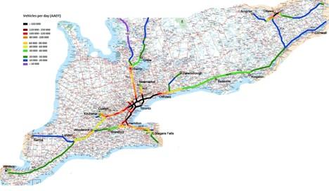
Imagine you’re driving from London to Toronto to visit family over the holidays. You decide to take Highway 403 since it is the quickest and most direct path there. Highways and roads act as long and short pathways to connect cities – similarly, different areas of our brains are connected with long and short cellular pathways. Signals are sent down these pathways which allows different regions of our brain to communicate with one another. This communication can be measured with different brain imaging techniques. For example, diffusion MRI measures the movement of water in the brain. Water moves more easily when flowing along cellular pathways, as opposed to when it has to move across them. By measuring this movement, we can find out where and also how many paths someone has in their brain. In the same way Toronto has more roadways which connect it to surrounding cities than the smaller city of Sarnia might, some regions are known to be very connected to the other areas in the brain, while others are known to have less connections.
https://cityjungles.wordpress.com/wp-content/uploads/2011/08/ontario_freeway.jpg
Another brain imaging technique used to measure communication is functional MRI. When an area of your brain is active, it requires oxygen. If two areas are using oxygen at the same time, we know that they are both active at the same time and communicating with one another. Functional MRI measures this flow of oxygen. Similarly to the way Toronto has more cars pass through it on a daily basis when compared to the less populous city of Sarnia, some brain areas are known to be more active than others.
Toronto has many roadways and many cars are able to pass through it – you might call it a hub of Ontario. We also use the term hub to refer to brain areas which tend to have many connections and are able to communicate with many other areas. In some brain disorders, such as epilepsy, this communication can break down.
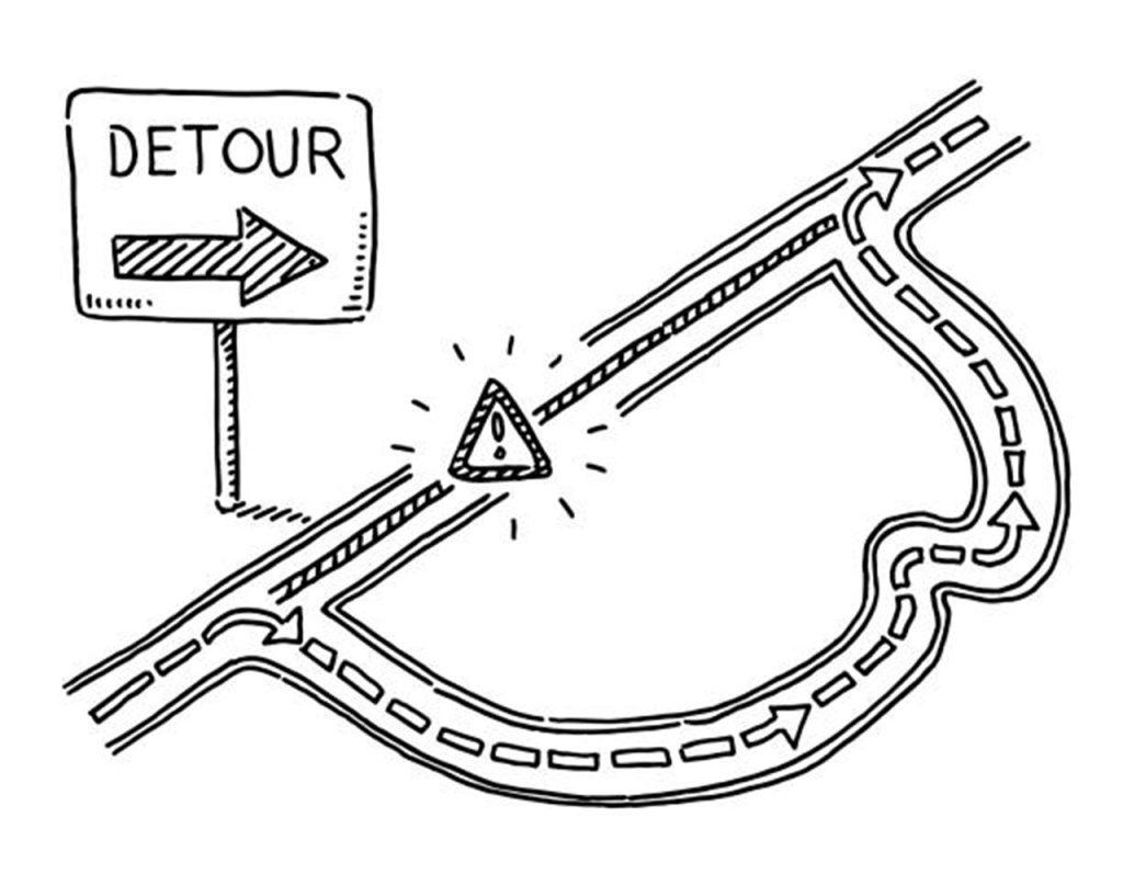
Think back to your journey over the holidays – halfway through your drive you see the dreaded road closed sign and are directed off the highway onto backroads to take a detour. You still reach your destination, but it takes you on a longer route, requiring more time. When pathways break down, our brains can also use detours. In epilepsy, long range “highway” pathways tend to be disrupted. In order to continue the flow of information, activity will be passed along the shorter “backroad” connections instead. Since brain hubs have so many connections, when any pathways breakdown the hubs are likely to be affected. In this way, studying how brain hubs in individuals with and without epilepsy are different might allow us to figure out how epilepsy affects communication in the brain.
Like roadway statistics, there are many different ways researchers can look at the communication of brain hubs. Instead of which direction and type of cars are driving down the roadways, we can analyze the flow of communication and what kind of information is being sent between hubs. There are also various imaging techniques which can be used. While this field is providing a countless amount of information about how the brains of individuals with epilepsy work, the best measures and techniques are still being debated. Due to this, it is important that researchers continue to study how brain hubs are affected in epilepsy. Once we understand how the brains of individuals with epilepsy are different, we can begin to better understand what kind of treatments might help them live a seizure free life.
Original article: Royer J, Bernhardt BC, Lariviere S, Gleichgerrcht E, Vorderwulbecke BJ, Vulliemoz S, Bonilha L. Epilepsy and brain network hubs. Epilepsia. 2022;63(3):537-550.
doi: 10.1111/epi.17171